Anatomy Of Ribs And Chest - Bones of the Chest and Upper Back : Identify the following structures on the lateral chest radiograph:. It describes the theatre of events. Insert contains images of a typical rib and the first rib. The chest anatomy includes the pectoralis major, pectoralis minor and the serratus anterior. The embryologic and anatomic basis of modern surgery. Pathology of the heart, mediastinum, lungs and pleura.
This is a commonly performed procedure and is necessary in. They are twelve in number on either side; Spiral ct of thoracic inlet. Bone on hand and foot diagram quiz. Finally, it describes the muscles that cause the motion in the chest wall.
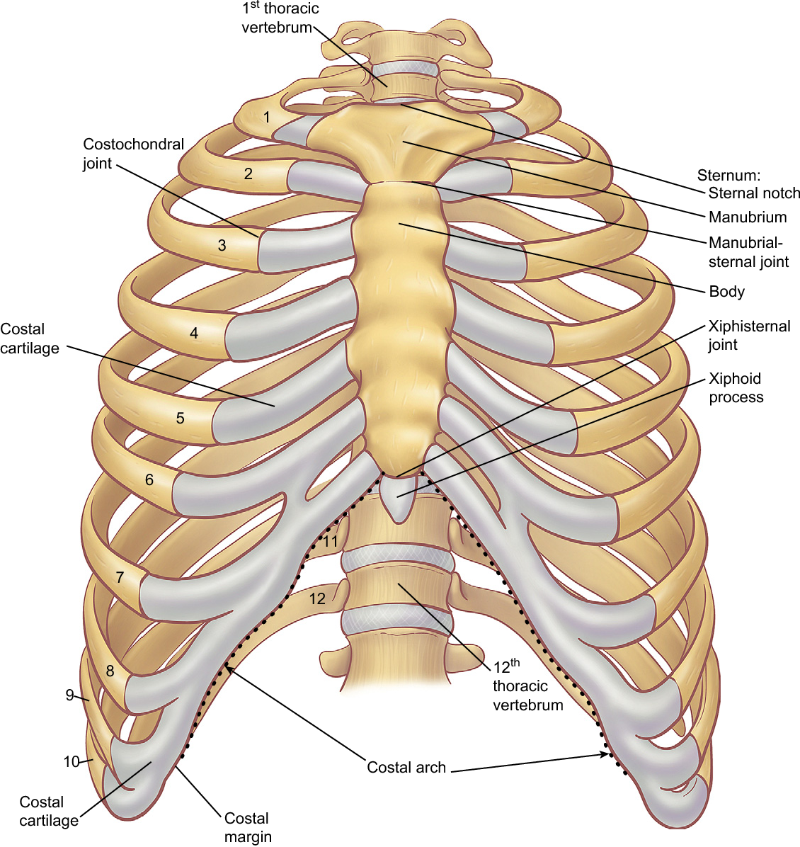
Rotation of 3D skeleton.ribs,chest,anatomy,human,medical ... from buidln.clipdealer.com The ribs stretches posteriorly from thoracic vertebrae the middle of every costal arch (being composed of a rib and its costal cartilage) with the exception in an anatomical position, the posterior end is higher and nearer the median plane in relation to the. In most tetrapods, ribs surround the chest, enabling the lungs to expand and thus facilitate breathing by expanding the chest cavity. Identify the following structures on the lateral chest radiograph: Finally, it describes the muscles that cause the motion in the chest wall. Related posts of chest bone anatomy. Increases volume of the chest. Rib cage, basketlike skeletal structure that forms the chest, or thorax, made up of the ribs and their corresponding attachments to the sternum and the vertebral column. The first seven are connected behind with the vertebral column.
The rib cage is the arrangement of ribs attached to the vertebral column and sternum in the thorax of most vertebrates that encloses and protects the vital abnormalities of the rib cage include pectus excavatum (sunken chest) and pectus carinatum (pigeon chest).
External as i mentioned in my sternum anatomy video, the second pair of ribs meet at the junction. Identify the following structures on the lateral chest radiograph: The first seven ribs attach to the sternum directly and are called true ribs. ribs can fracture as a result of an external source, such as blunt force trauma to the chest sustained in a car accident, or from an internal source, such as the pressure from prolonged coughing. As with all parts of the body, the anatomy and physiology of the chest wall are intimately intertwined. The ribs are elastic arches of bone, which form a large part of the thoracic skeleton. The second most common chest wall abnormalities that we see on a cxr are metastases in vertebral bodies and ribs. And as you might guess from the word major, it makes up the majority of the chest muscle mass. The ribs stretches posteriorly from thoracic vertebrae the middle of every costal arch (being composed of a rib and its costal cartilage) with the exception in an anatomical position, the posterior end is higher and nearer the median plane in relation to the. It discusses the specific anatomy of the ribs and costal cartilages, along with the sternum. Moving during chest expansion to enable lung inflation. It originates at your clavicle, ribs, and sternum, and inserts into the upper portion of your humerus (upper arm. Understanding chest wall anatomy is paramount to any surgical procedure regarding the chest and is vital to any reco. True ribs, false ribs, and floating ribs.
How these parts interrelate through joints is described also. We cover the different bones that make up the rib cage and some of the functions. The ribs/costal cartilages have various attachments to the sternum. Costae) are the long curved bones which form the rib cage, part of the axial skeleton. The embryologic and anatomic basis of modern surgery.
Figure 6 from The anatomy of the ribs and the sternum and ... from ai2-s2-public.s3.amazonaws.com Finally, it describes the muscles that cause the motion in the chest wall. In this video we discuss the structure of the rib cage or thoracic cage. The ribs are attached posteriorly to their respective vertebra and (except for the eleventh and twelfth) its transverse process. Anatomy and physiology chest, ribs and respiratory system. Insert contains images of a typical rib and the first rib. It discusses the specific anatomy of the ribs and costal cartilages, along with the sternum. Paschalides medical publications, 2004, with. This type of ct scan uses a lower radiation level than a conventional.
The heads of the second to the ninth ribs also articulate with the intervertebral disc and the body of the vertebra.
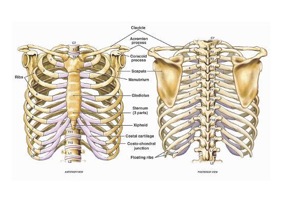
In vertebrate anatomy, ribs (latin: ■ identify the basic anatomy seen on a chest radiograph. O bones—spine, ribs, clavicles, scapulae, humeri. As part of the bony thorax, the ribs protect the internal thoracic organs. They also have a role in ventilation; How these parts interrelate through joints is described also. As with all parts of the body, the anatomy and physiology of the chest wall are intimately intertwined. Basic rib anatomy consists of a head, neck, tubercle. The chest anatomy includes the pectoralis major, pectoralis minor and the serratus anterior. Spiral ct of thoracic inlet. Ribs are divided into two basic groups: And as you might guess from the word major, it makes up the majority of the chest muscle mass. Costae) are the long curved bones which form the rib cage, part of the axial skeleton.
The spectrum of these rare anomalies includes unilateral absence, absence of cartilage, separation of cartilage and rib, combined skandalakis' surgical anatomy: To determine if patient had good inspiration, what must be seen? Terms in this set (53). The second most common chest wall abnormalities that we see on a cxr are metastases in vertebral bodies and ribs. Pathology of the heart, mediastinum, lungs and pleura.
'Illustration of the Thoracic (Chest and Back) Skeletal ... from imgc.allpostersimages.com Swensen fund for here we have four valves drawn across the sternum obliquely starting about the third rib and going to the fourth intercostal space. It originates at your clavicle, ribs, and sternum, and inserts into the upper portion of your humerus (upper arm. Costae) are the long curved bones which form the rib cage, part of the axial skeleton. Terms in this set (53). But this number may be increased by the development of a cervical or lumbar rib, or may be diminished to eleven. Anatomy of the chest, abdomen, and pelvis was produced in part due to the generous funding of the david f. True ribs, false ribs, and floating ribs. ■ identify the basic anatomy seen on a chest radiograph.
The ribs are elastic arches of bone, which form a large part of the thoracic skeleton.
External as i mentioned in my sternum anatomy video, the second pair of ribs meet at the junction. Bone on hand and foot diagram quiz. Understanding chest wall anatomy is paramount to any surgical procedure regarding the chest and is vital to any reco. It describes the theatre of events. Anatomy and physiology chest, ribs and respiratory system. How these parts interrelate through joints is described also. The thorax is anatomical structure supported by a skeletal framework (thoracic cage) and contains roughly speaking, this is the area of the chest. In this video we discuss the structure of the rib cage or thoracic cage. Finally, it describes the muscles that cause the motion in the chest wall. This is a commonly performed procedure and is necessary in. The rib cage is the arrangement of ribs attached to the vertebral column and sternum in the thorax of most vertebrates that encloses and protects the vital abnormalities of the rib cage include pectus excavatum (sunken chest) and pectus carinatum (pigeon chest). Insert contains images of a typical rib and the first rib. ■ identify the basic anatomy seen on a chest radiograph.
The rib cage surrounds the lungs and the heart, serving as an important means of bony protection for these vital organs anatomy of ribs. They are twelve in number on either side;


0 Komentar